

Why Are Microscopes Used in Hair Transplant Surgery?
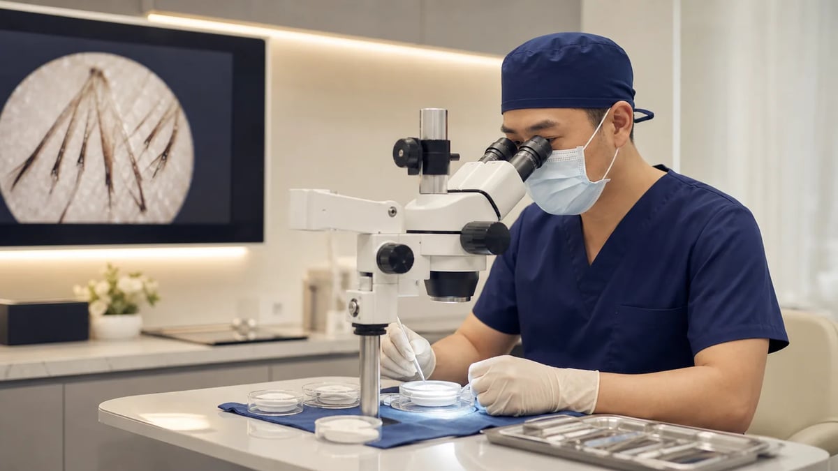
A hair transplant is not simply moving hair from the back of the scalp to a thin area. After extraction, grafts must be inspected, prepared, classified, stored, and implanted. Magnification helps a trained team see follicular structures that are difficult to judge with the naked eye and reduce avoidable pulling or cutting.
Key point: a stereomicroscope is an important graft-handling tool, but no machine determines the result by itself. Graft viability and naturalness also depend on harvesting, handling, time outside the body, implantation direction, donor quality, and aftercare.
If you are considering a hair transplant, look beyond the labels FUE and FUT. Ask how extracted grafts are handled and who is responsible for diagnosis, hairline design, and the critical surgical steps.
What is a follicular unit?
Scalp hair commonly grows in natural follicular units rather than as completely separate strands. A unit may contain one or several hairs along with supporting tissue. Modern transplantation aims to preserve these natural groupings and place suitable units in the right parts of the recipient area.
Single-hair grafts are often selected for the leading edge of a hairline, while multi-hair units can add visual density behind it. This is a design decision; cutting every graft into the smallest possible piece is not the goal.
| Handling step | Purpose | Main caution |
|---|---|---|
| Identify follicular units | Preserve natural groupings | Avoid injury to the shaft, bulb, and supporting structures |
| Trim excess tissue | Prepare a graft for placement | Avoid over-trimming and dehydration |
| Classify by hair count | Match hairline and density design | Classification must follow the overall plan |
| Store outside the body | Keep grafts hydrated and usable | Time, temperature, and solution require a controlled protocol |
What does the microscope do during a hair transplant?
A stereomicroscope enlarges the direction of follicles, bulbs, and surrounding tissue so trained staff can separate, inspect, and classify grafts more precisely. The American Academy of Dermatology also explains that technicians prepare harvested hairs before the surgeon readies the recipient scalp and transplantation proceeds.

Its role differs by technique. In FUT, a donor strip is divided into follicular units under magnification. In FUE, units are removed individually, but extracted grafts may still need inspection, minimal trimming, or classification. Not every FUE graft should be split again.
Can dissection damage a follicle?
Any manipulation can cause mechanical injury. The objective is therefore to see the structures clearly and perform only the preparation that is necessary—not to make every graft as small as possible. A comparative study reported poorer survival among grafts with several forms of injury, with fractured follicles performing particularly poorly.
This is why quality cannot be judged only by the punch or implanter brand. Harvest angle, forceps handling, microscopic preparation, counting, transfer, and implantation are parts of one workflow. Compare the broader trade-offs in our FUE, FUT, and COMBO guide.
Why do storage and time outside the body matter?
Once removed, a graft temporarily loses its normal blood supply. It needs to remain hydrated and protected from unnecessary compression, drying, and delay. Storage solution, temperature, and out-of-body time should be managed through a clinical protocol.

Research continues to compare storage conditions, so a single laboratory result should not be treated as a universal rule for every operation. A practical surgical workflow uses traceable batches and coordinated harvesting and implantation to avoid needless exposure. Patients do not need to prescribe a particular solution, but they can ask how the team manages the grafts.
How can microscopic classification affect a natural hairline?
A natural hairline usually transitions from a fine, slightly irregular leading edge into greater density behind it. Classification helps the surgeon reserve suitable single-hair grafts for the front and use multi-hair units farther back. The surgeon must still decide the angle, direction, spacing, and overall proportions.
The microscope supports “seeing and preparing”; diagnosis and aesthetic planning determine “where, which direction, and how many.” Neither replaces the other.
What should you ask about the doctor and surgical team?
Instead of asking only which device the clinic uses, check whether the diagnosis, donor assessment, division of responsibilities, and risk discussion are clear:
- Does the doctor assess the cause and likely future pattern of hair loss?
- Are the benefits and limitations of FUE, FUT, and alternatives explained?
- Is there a consistent process for graft inspection, storage, classification, and counting?
- Are hairline angle, direction, and density planned for the individual?
- Is there a clear route for aftercare and urgent questions?
You can also read Should you choose a hair-transplant clinic or surgeon? and our graft survival and failure guide.
This article provides general medical education and cannot replace an individual consultation. Suitability, technique, and expected results must be assessed from the diagnosis, donor supply, health, and long-term plan.
References
Does every hair transplant graft need to be dissected? +
No. FUT requires strip tissue to be divided into follicular units. FUE grafts are already removed one by one and are usually inspected, minimally trimmed, and classified; further splitting depends on graft structure and hairline planning.
Does higher microscope magnification guarantee a better result? +
No. Magnification helps the team see graft anatomy, but outcomes also depend on training, harvesting, implantation, storage, design, donor quality, and healing.
Are single-hair grafts always more natural? +
They are often useful at the leading edge of a hairline. Multi-hair units are still valuable behind it for visual density, so naturalness comes from the overall plan.
How long can grafts remain outside the body? +
There is no single safe number for every procedure and storage protocol. Grafts should stay hydrated, be protected from unnecessary trauma, and avoid needless waiting.
Are microscopes used with FUE as well as FUT? +
They can be. FUE grafts may be inspected for integrity, trimmed, and classified under magnification, although this is different from dissecting an FUT strip.
How can a patient assess a clinic's graft-handling process? +
Ask who inspects and classifies grafts, how they are kept hydrated, how counts are checked, and who provides follow-up. The clinic should explain the workflow clearly.
Related articles
This article has been reviewed and medically approved by Dr. Wen-Yi Wu

Dr. Wen-Yi Wu|Director, Mong Hair Clinic
- ● Fellow of ISHRS (FISHRS)
- ● ABHRS Board-Certified Hair Restoration Surgeon
- ● President of TSHRS (Taiwan Society of Hair Restoration Surgery)