

One-Year Hair Transplant Review: Density, Direction and Native Hair
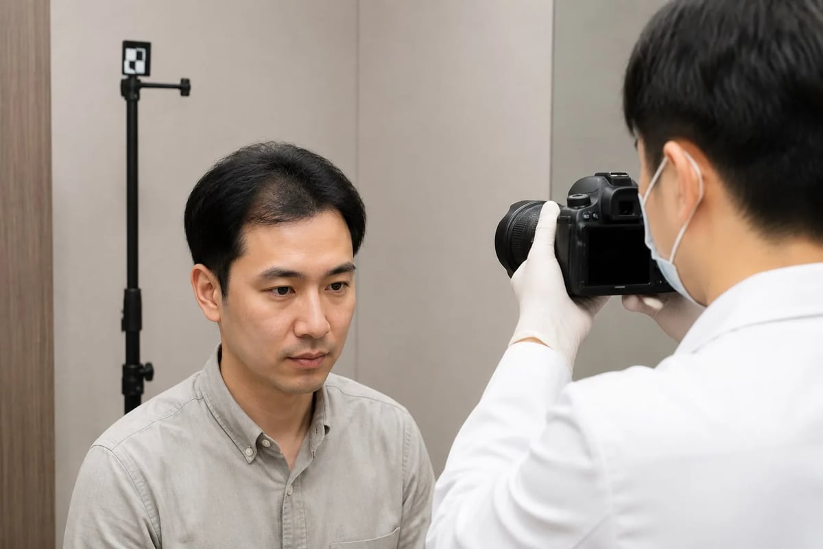
A hair transplant needs time before the follicles enter a new growth cycle. At the one-year review, the question is not simply whether hair has appeared. A doctor should compare the original plan, surgical record and photographs taken under consistent conditions, then assess recipient-site coverage, hair calibre and direction as well as changes in native hair and the donor area.
One year is a meaningful follow-up point, but it is not a universal deadline. Hair length, lighting, strand thickness, camera angle and continuing miniaturisation of nearby native hair can all change the apparent density.
Key point: a one-year review should assess standardised photographs, the recipient area, native hair and the donor area together. If density seems lower than expected, establish the reason before discussing observation, medical treatment or another procedure. A selfie or planned graft count cannot determine success on its own.
Why is one year a useful time for a full review?
Transplanted follicles do not show a mature appearance immediately. Hair shafts may shed before follicles rest and grow again, while new hairs gradually gain length and calibre. Around one year, everyday coverage at the hairline, parting or crown can usually be assessed more meaningfully than it can in the early months.
However, “one year” is a review point rather than an identical endpoint for every patient. The treated area, individual growth cycle, native-hair condition and postoperative health can affect maturation. For a stage-by-stage overview, see the hair transplant growth timeline; this article focuses on how to judge the one-year review.
What records should be compared at the one-year review?
A useful review involves more than placing a preoperative image beside a current selfie. Camera angle, wet or dry hair, length, parting and light source can substantially change scalp visibility. A more reliable comparison uses similar frontal, side, top and donor-area views alongside the original design.
| Record | What it helps assess | Main limitation |
|---|---|---|
| Standard preoperative photographs | Original hairline, thinning pattern and donor appearance | Different angles, lighting and length prevent direct comparison |
| Surgical record | Technique, treated area, planned graft count and harvesting method | Planned grafts are not the same as hairs that can be counted in a photograph |
| Current multi-angle photographs | Coverage, symmetry, parting and crown pattern | Wet hair, styling products and strong overhead light change apparent density |
| Medicine and health changes | Native-hair progression, shedding and other contributors | These require clinical history and scalp examination |
If surgery was performed elsewhere, bring the operation date, technique, treated area, estimated graft count, preoperative photographs, medicines and any postoperative concerns. A current examination is still possible without a complete record, but the comparison with baseline will be less precise.
Should the recipient area be judged by density, calibre or direction?
All three matter, and hair count alone is not enough. The same number of follicles can provide different visual coverage when strands are fine or strongly contrast with the scalp. Direction that does not blend with native hair may also become more noticeable with longer hair or a particular parting.

| Observation | What the doctor examines | What it cannot show alone |
|---|---|---|
| Visual density | Scalp coverage and hairstyle at an everyday viewing distance | It cannot precisely reconstruct graft survival |
| Hair calibre | Variation in strand thickness and the presence of fine hairs | A fine hair does not necessarily mean a non-functioning follicle |
| Direction and angle | How growth blends with the design and surrounding hair | One strand cannot represent the naturalness of an entire area |
| Distribution | Obvious gaps, unevenness or the influence of scarring | Different zones may intentionally use different densities |
Surgical records commonly use follicular units or grafts, and each unit may contain a different number of hairs. A photograph cannot verify every graft. If the main concern is survival, read our guide to hair transplant survival and failure assessment.
Why does a thinner-looking photograph not necessarily mean failure?
Strong overhead light, low-angle flash, wet or oily hair, a shorter haircut and a wider part can expose more scalp. Soft light, volume and longer hair can make coverage appear greater. This is why repeatable photography matters.
The photograph also contains both transplanted and native hair. Pattern hair loss can continue to miniaturise native hairs around or behind the transplant. A reduction in overall visual density does not automatically mean the transplanted follicles stopped growing. When comparable photographs still show a clear local difference, the doctor can assess calibre, distribution, scalp disease, inflammation and scarring.
Why should native and transplanted hair be assessed separately?
A hair transplant redistributes donor follicles; it does not stop the progression of pattern hair loss. Transplanted hairs and the native hairs retained in the recipient area have different histories. Looking only at the combined photograph can make native-hair miniaturisation look like poor transplant growth.
For a fuller explanation, see how transplanted and native hair can change after surgery. Finasteride, minoxidil or another treatment should be considered individually according to diagnosis, health, contraindications and preference. Do not start or stop prescription treatment simply to influence a follow-up image.
What should be checked in the donor area after one year?
The frontal result is only part of the review. After FUE, the doctor should assess extraction distribution, remaining density, hair calibre and visibility with shorter hair. After FUT, the linear scar, flexibility and coverage by surrounding hair should be examined. In either technique, the remaining long-term donor reserve matters.
An even-looking donor area does not mean the extracted follicles regenerated in their original sites; surrounding retained hair usually supplies the coverage. See our guide to FUE donor-area regrowth and thinning risk.
Which symptoms need an earlier review?
Increasing redness, pain, bleeding, pustules, discharge, wound separation, skin-colour change or expanding numbness should not be managed by waiting for the one-year appointment. Contact the operating clinic or another appropriately qualified doctor promptly.
Sudden extensive shedding, a visibly patchy donor area or recurrent scalp inflammation also warrant assessment. Fever, rapidly spreading redness, severe pain or systemic illness needs urgent medical attention.
Can limited density at one year be treated with another transplant immediately?
Not automatically. Before a second procedure, the doctor needs to decide whether the current growth is ready to judge, where the limitation lies, whether native hair is still miniaturising and whether the donor area can be harvested safely. If the concern mainly reflects lighting, hairstyle or changing native hair, immediate additional grafting may not be the best response.

| Situation at one year | What to clarify first | Possible next step |
|---|---|---|
| Thin appearance only under certain lighting | Photography, length, parting and calibre | Repeat standardised photographs and examine the scalp |
| Continuing thinning around the transplant | Native-hair progression, medicines and other causes | Diagnose first, then discuss individual treatment |
| Local direction or distribution concern | Existing flow, scar and realistic adjustment range | Compare styling options with revision risks |
| Redness, itching or papules | Inflammation, folliculitis or another scalp disorder | Treat the medical problem before adding density |
| Confirmed need for more coverage | Donor reserve, future loss and procedural risk | Plan whether and when a second procedure is reasonable |
Donor follicles are finite. Read more about how many hair transplants may be possible and how repeat surgery is assessed.
How should I prepare for the review?
Bring photographs from before surgery and different postoperative stages, the surgical record if available, current medicines and a list of symptoms. Attend with clean hair in its usual state and avoid hair fibres, scalp concealers or heavy styling products that obscure the skin.
Frame concerns as specific situations—such as a part that remains visible under overhead light, one side that is difficult to style, or a donor patch that shows after a short haircut. This gives the doctor a more useful starting point than the single question, “Was it successful?”
Dr Wen-Yi Wu: the one-year review is more than checking for growth
I compare the original design and surgical record with photographs taken under consistent conditions, then examine the recipient area, native hair and donor area separately. If the appearance differs from expectations, we first clarify lighting, calibre, direction and native-hair change before deciding whether observation, treatment or revision is appropriate.
At Taiwan Hair, Dr Wen-Yi Wu provides the medical consultation, surgical planning and follow-up assessment directly rather than delegating medical decisions to a non-medical sales consultant. This article provides general medical information and cannot replace an individual diagnosis or outcome assessment.
References
- Hair transplantation: standard guidelines of care
- Hair Transplant Practice Guidelines
- Donor-site appearance after follicular unit excision
- Standardized photography and follow-up in FUE outcome assessment
- Standard Operating Procedures in Trichological Practice
- A Scoping Review on Complications in Modern Hair Transplantation
Can transplanted hair continue to change after one year? +
One year is a useful overall review point, but growth and maturation vary. Some hairs may still change in calibre, length or appearance. The doctor should consider the treated area, standardised photographs and scalp examination before drawing a conclusion.
Why do clinic photographs look different from my selfies? +
Lighting direction, camera angle, distance, hair length, parting, wetness and styling products all change how much scalp is visible. Compare photographs taken under similar conditions and combine them with an examination.
Does visible scalp at one year mean the transplant failed? +
Not necessarily. Visual coverage also depends on hair calibre, hair-to-skin colour contrast, distribution, direction and changes in native hair. The recipient area, native hair and surgical record need to be reviewed together.
Should I stop hair-loss medicine before the review? +
Do not stop treatment simply to change the review photographs. Starting, changing or stopping finasteride, minoxidil or another treatment should be discussed with a doctor who knows your diagnosis and health history.
Can the crown and frontal hairline be judged in the same way? +
Not from one angle or hairstyle. The two areas have different flow patterns, lighting and coverage characteristics, and they may mature at different rates. Each area should be compared with its original plan.
When can a second transplant be considered if density is limited at one year? +
There is no single interval for everyone. The doctor should first determine whether growth is ready to assess, whether native hair is stable, and whether the scalp, scars and donor reserve make another procedure appropriate.
Related articles
This article has been reviewed and medically approved by Dr. Wen-Yi Wu

Dr. Wen-Yi Wu|Director, Mong Hair Clinic
- ● Fellow of ISHRS (FISHRS)
- ● ABHRS Board-Certified Hair Restoration Surgeon
- ● President of TSHRS (Taiwan Society of Hair Restoration Surgery)